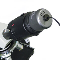
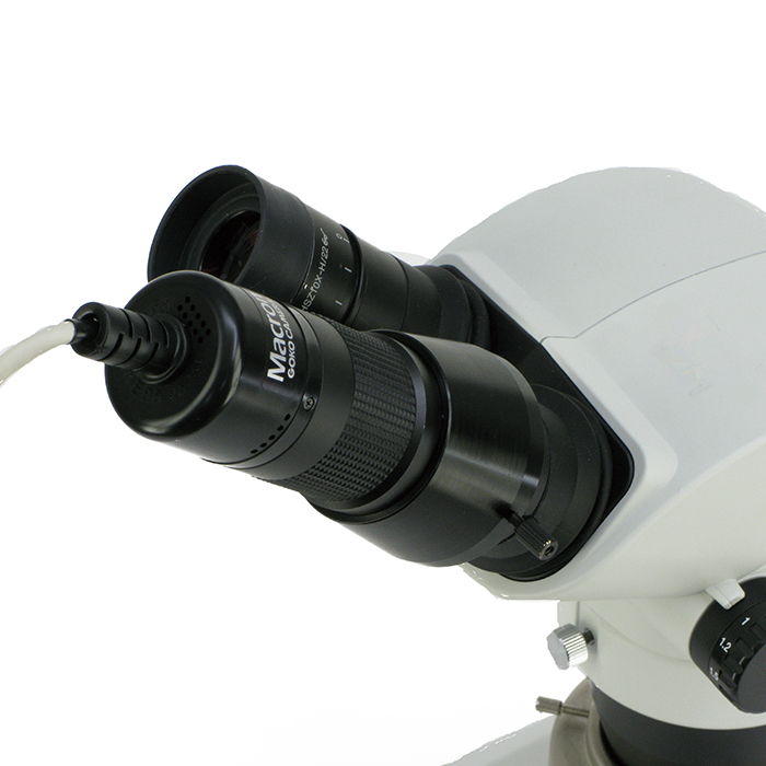
Upgrades your optical microscope to a digital microscope.
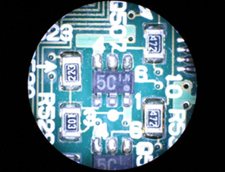
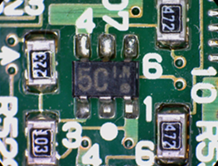
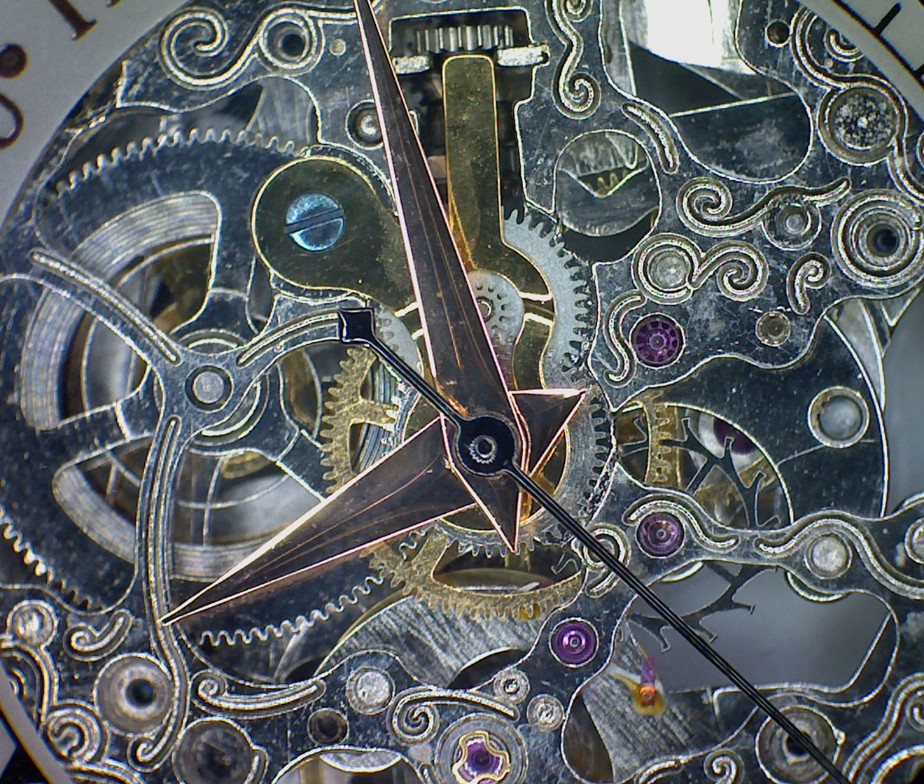
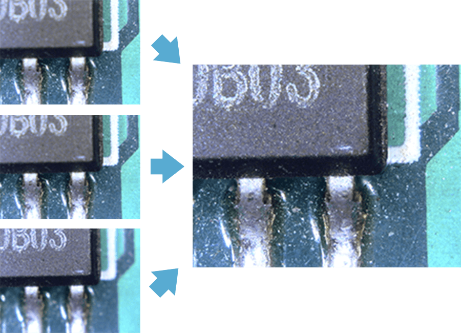
Offers a larger image field that is close in size to the eyepiece view
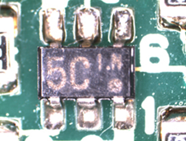
- Praised for clarity, ease of use, and automatic exposure control.
- UVC-compatible (no driver needed); works with Windows 10/11, Type-C iPads, Macs and Android. (units sold after July 2016)
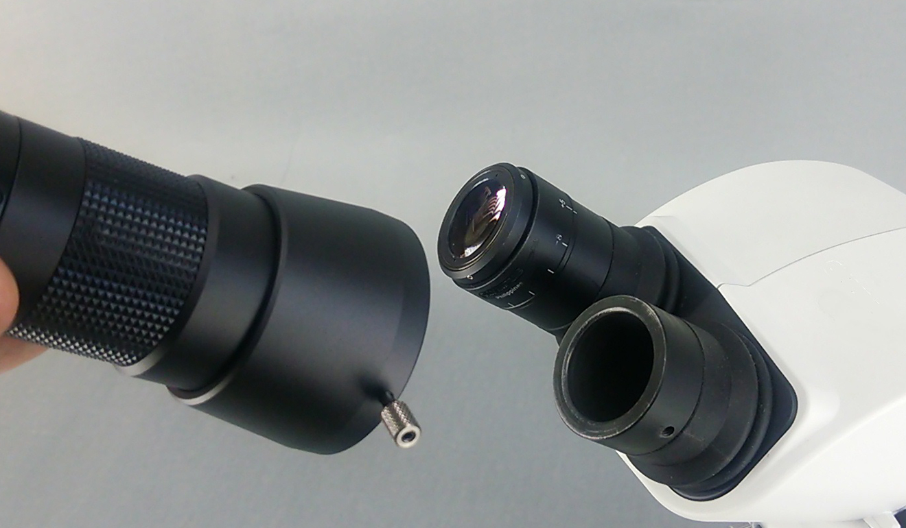
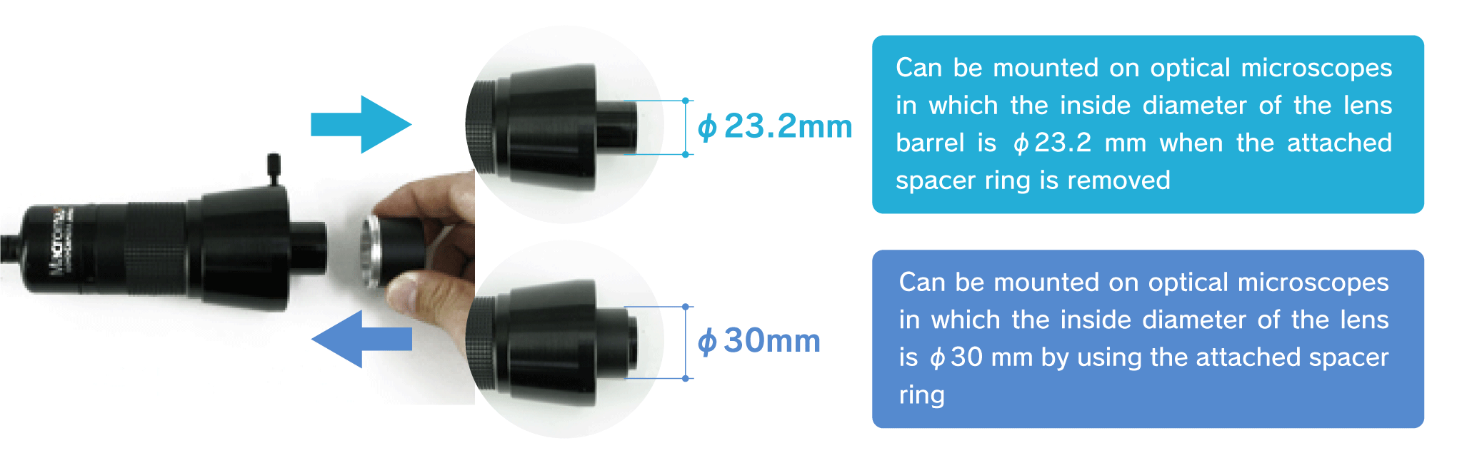
- Fits ISO-standard 23.2mm / 30mm eyepiece tubes (Olympus, Nikon, Leica, ZEISS, etc.), including phase-contrast and metallurgical microscopes.
- No trinocular tube? No problem. Mounts easily on standard eyepiece tubes.
- Hassle-free attachment and removal with a user-friendly design.
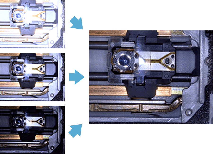
- Wide field of view―about 3-5× larger than conventional models―similar to the naked-eye view.
- Includes free measurement software with auto edge detection to reduce user error; choose free or premium version.
- Trusted by QC departments, research institutes, and universities.
- Affordable, reliable, and made in Japan with GOKO's full support.